
Contact Penta
+49 94 35 30 71 38
info (at)penta-arzneimittel.de
Anatomy of the respiratory tract
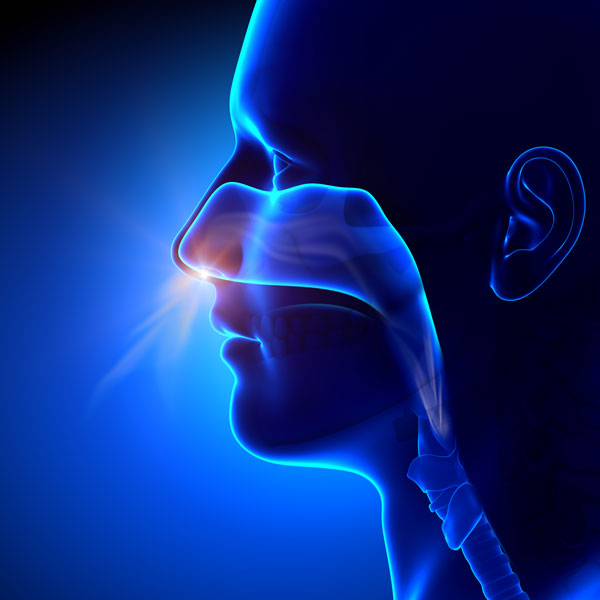
Upper respiratory tract - nose, mouth and throat
Tasks of the respiratory system
The respiratory tract is not only used to supply fresh air during inspiration (= inhalation) and to exhaust "alveolar air" during expiration (= exhalation), but it also fulfils a number of auxiliary functions to aid breathing. These include cleansing, warming up and humidifying the inhaled air.
The inhaled air is partly purified in the nose, where smaller particles, dust and bacteria are trapped by the mucous membranes (this is why chronic breathing through the mouth increases susceptibility to respiratory diseases).
Other inhaled particles are deposited on the layer of mucus covering the walls of the incoming airways.
(Inhaled air is routed through the nasopharynx, the larynx and its narrowest point, the glottis, into the trachea (windpipe). The trachea is a large-lumen tube of connective tissue, which forks, at the level of the 5th thoracic vertebra, into the two main bronchial tubes (stem bronchi), which in turn each branch into one of the lobes of the lung below.)
The mucus secreted by goblet cells and subepithelial gland cells is constantly transported towards the mouth via rhythmic movement of the cilia of the respiratory epithelium and then swallowed. Mucus transport therefore ensures that foreign particles and bacteria are removed from the respiratory tract. If the cilia are damaged, as is the case with chronic bronchitis, for example, mucus accumulates in the respiratory tract, leading to increased respiratory resistance.
Lower respiratory tract - bronchial system
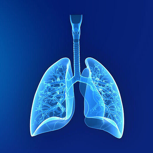
Anatomy of the bronchial system
The air-conducting (conductive) part is solely responsible for the transport of the inhaled and exhaled air. It is part of the anatomical dead space of the bronchial system. It includes the trachea, bronchi and bronchioli lobulares and terminales. The respiratory section consists of the bronchioli respiratorii and the ductus alveolares with their air sacs (alveoli). It corresponds to the portion of the bronchial tree which is involved in gas exchange.
Anatomy of the lungs
The lung consists of two separate lobes which fill the lateral halves of the thorax area on both sides. Their outer surfaces rest against the inner thoracic wall, while their lower surfaces rest on the diaphragm. Located between the two lobes of the lung are the organs, of which the heart occupies the largest space. Each lobe is covered by a sheath supplied with vessels (pleura visceralis).
Function of the alveoli
The exchange of respiratory gases between the gas-filled cavities and the blood in the lung capillaries takes place in the area of the air sacs (= alveoli). The alveoli measure about 0.2 mm in diameter; their number is estimated at about 300 million and their total surface at about 80 m². They are surrounded by a dense capillary network.